High-Resolution Macromolecular Cryo-EM Facility
Equipped with leading-edge technology, HRMEM provides instrumentation, training and consultation for both single particle and cryo electron tomography TEM workflows, and support academics across institutions and dozens of industry partners.
Instrumentation includes:
- ThermoFisher Talos 120kV microscope with a Ceta camera
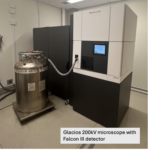
- ThermoFisher Glacios 200kV microscope has a Falcon III detector
- FEI Titan Krios G2 300kV with a ThermoFisher Selectris energy filter with a Falcon 4i detector
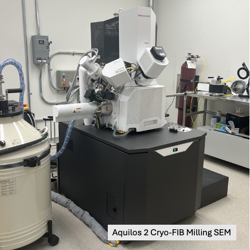
- Aquilos 2 Cryo-FIB Milling SEM and iFLM arm
- Leica Stellaris 5 cryo CLEM
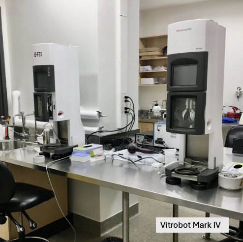
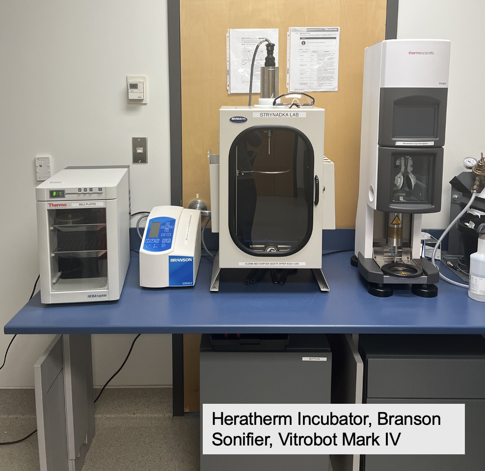
- Two Vitrobot Mark IV robots
- Leica EM GP Automatic plunge freezer with bare grid technique
- Solarus II Plasma Cleaner
- Leica ACE200 Sputter Coaters for Au and Carbon Grid fabrication


Bio-analytical and Binding Suite
Equipped with state-of-the-art instrumentation, the Bio-Analytical and Binding Suite enables researchers to characterize the kinetic and thermodynamic parameters of protein–protein and protein–ligand interactions. It supports detailed analysis of sample properties such as stoichiometry, monodispersity, and stability through techniques like DLS, MALS, ITC, and SPR.
Instrumentation includes:
- Refeyn Mass Photometer
- Agilent Synergy H1 Hybrid Multi-Mode Reader
- BioTek Synergy H4 Microplate Reader
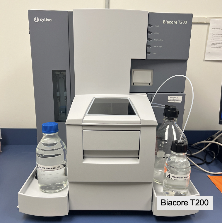
- Biacore T200 SPR System (Surface Plasmon Resonance)
- MicroCal PEAQ-ITC Microcalorimeter (Isothermal titration calorimetry ITC)
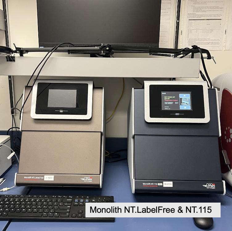
- Monolith NT.115 PICO Microthermophoresis (MST) unit
- Monolith NT.LabelFree Microthermophoresis unit
- Octet Red96 BLI Detection System (Biolayter interferometry)
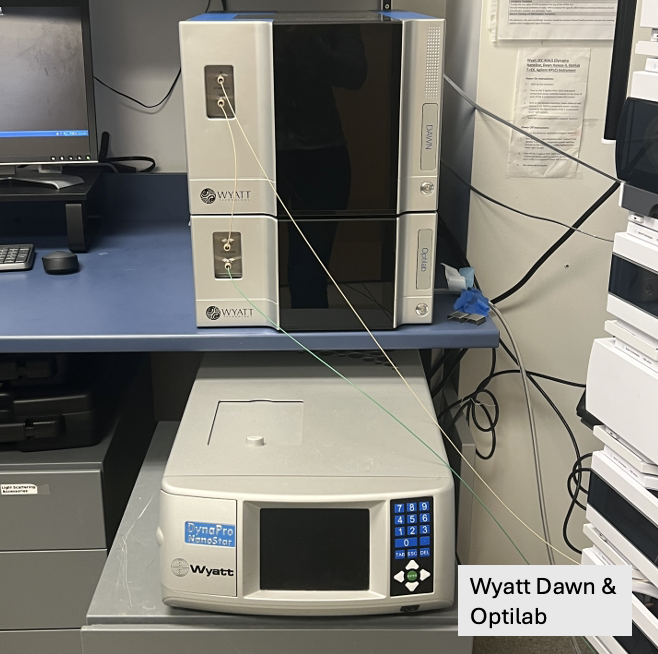
- Wyatt Optilab Multiwavelength Static Light Scattering (MALS)
- Wyatt DynaPro NanoStar (Dynamic Light Scattering DLS)
- Stargazer Differential Static Light Scattering with temperature gradient
- Microvolume ultracentrifuge


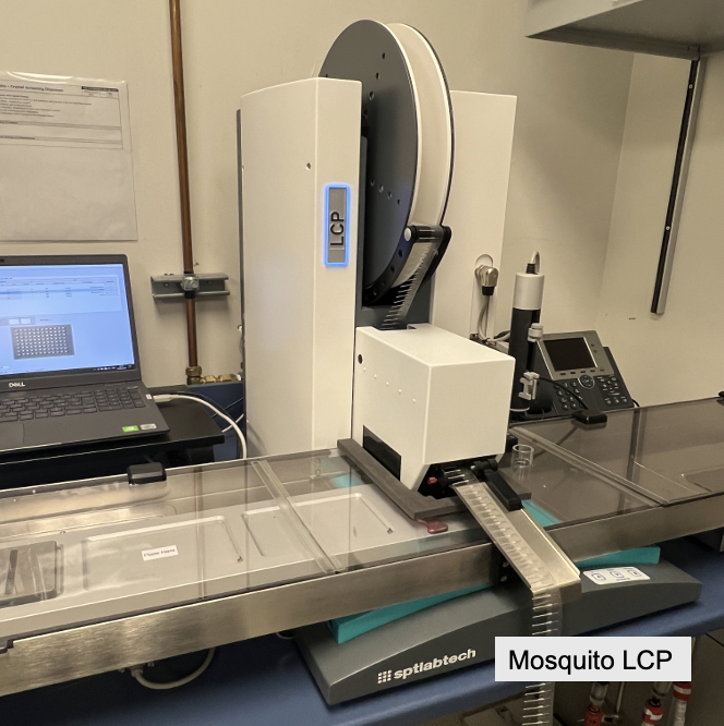
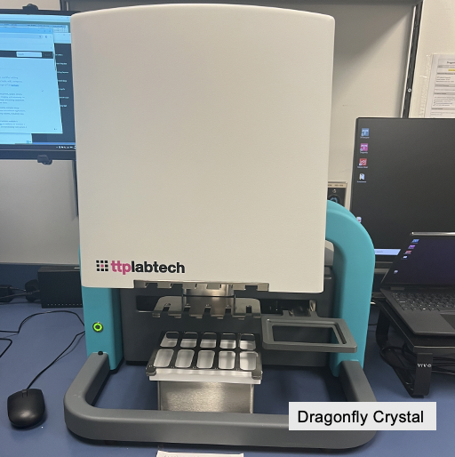
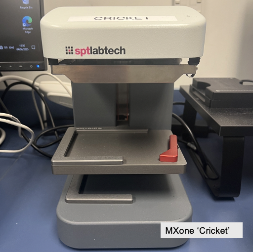
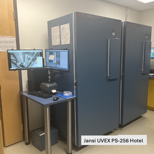
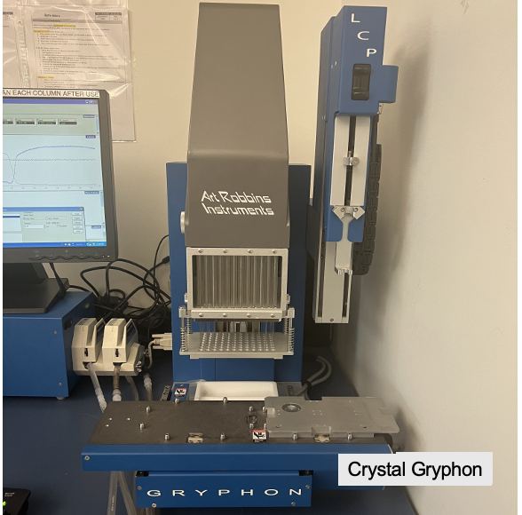
Robotics Crystallization Suite
The Robotics Crystallization Suite offers specialized instrumentation to support macromolecular crystallization workflows. The facility includes automated nanovolume sitting drop and lipidic cubic phase systems, customized screen generation robotics, temperature-controlled storage, and integrated UV imaging.
The suite is equipped with :

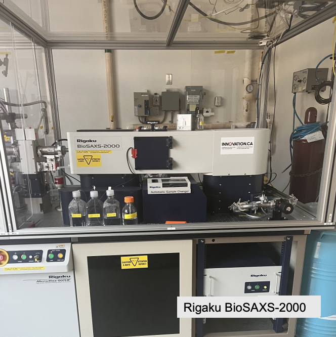
X-Ray Diffraction Facility
The X-Ray Diffraction Unit provides advanced capabilities for macromolecular structure determination using crystallographic methods. The facility includes a microfocus rotating CuKa anode generator with precision optics and modern detectors, along with automated sample handling and temperature control.
Instrumentation includes:
- Microfocus Rotating Anode X-ray Generator (MicroMax-007 HF Microfocus[MOU1] )
- With AFC11 goniometer, Dectris PILATUS3 R 200K Hybrid Pixel Array Detector and VM VHF Optics
- Haskris LX2 Water Chiller
- 800 Series Cryostream Controller
- Rigaku BioSAXS-2000 2D Kratky Small Angle X-Ray Scattering system
- Rigaku 96-well automatic sample changer

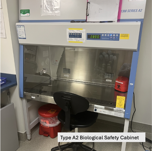
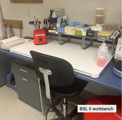
Biosafety Level II Suite
The certified Biosafety Level II protein production suite is designed for work with BSL II bacterial cultures (with each species subject to specific biosafety permits), enabling native expression of diverse bacterial targets.
Instrumentation includes:


NMR Spectroscopy Unit
Equipped with 600 and high-field 850 MHz Ascend spectrometers featuring cryoprobes, the NMR Suite enables high-resolution, sensitive analysis of protein structure, dynamics, and interactions. Its advanced instrumentation supports multidimensional experiments ideal for studying large proteins and complexes, ligand binding, conformational changes, and biomolecular assemblies.
Instrumentation includes:
- 600 and 850 MHz NMR spectrometers with cryoprobes

SFU Cellular Metabolite Sensing Unit
The King Lab at SFU has developed novel techniques for the assessment of microbial cellular metabolism by studying metabolite-responsive post-translational modifications (PTMs).
These modifications are assessed using:
- Chemoproteomic method to identify lysine residues modified by carbon dioxide (King et al., 2022)
- Proteome-wide approach to monitor the thermostability effect of PTMs (King et al., 2022)
Cobb Lab Nanobody Production
ASTRID collaborator Jennifer Cobb at the University of Victoria hosts a dedicated platform for nanobody discovery, engineering, and production, supporting applications in structural biology, therapeutic development, and advanced imaging. Services span the full pipeline from antigen preparation and yeast display screening to recombinant expression, purification, and optimization.
Expertise includes:
- Library construction, screening, and affinity maturation of nanobodies using yeast display
- Affinity testing with Biolayer Interferometry technology
- Recombinant expression and purification of final product

Cherkasov Lab Deep Docking Drug Discovery
The Cherkasov Lab at the Vancouver Prostate Centre, part of the ASTRID network of collaborators, hosts a dedicated platform for AI-driven drug discovery, supporting applications in infectious disease, oncology, and neurological disorders. The platform integrates structure-based design with machine learning to accelerate early-stage therapeutic development using ultra-large virtual screening.
Expertise includes:
- AI-powered virtual screening of ultra-large chemical libraries using the Deep Docking platform
- Structure-based hit identification, molecular docking, and cheminformatics analysis
- Access to high-performance computing infrastructure enabling scalable, rapid screening campaigns

Affiliated Research Hubs
UVic-Genome BC Proteomics Centre
The UVic-Genome BC Proteomics Centre is a hub for high throughput proteomics and metabolomics. The Centre offers protein structure characterization by hydrogen deuterium exchange mass spectrometry (HDX-MS)and cross-linkers, as well as metabolomics services and a variety of strategies for protein identification and quantification.
Expertise includes:
- Hydrogen deuterium exchange mass spectrometry (HDX-MS)
- Ultra-high pressure liquid chromatography (UPLC) and triple quadrupole mass spectrometers
- Matrix-Assisted Laser Desorption/Ionization Mass Spectrometry Imaging
- LC-MS/MS using Thermo Orbitrap mass spectrometers

LSI Proteomics Core Facility
The LSI Proteomics Core Facility offers mass spectrometry services for protein and peptide analysis, including mass confirmation, protein identification, post-translational modification profiling, and quantitative proteomics. The facility supports high-throughput workflows and provides expert consultation for experiment design and data interpretation.
Instrumentation Includes:
- Bruker Impact II
- Thermo Electron LTQ-Orbitrap VE
- Agilent 6550 QTOF
- Agilent 6460 QQQ
- Waters Xevo G2 QTOF
- Agilent 1200 HPLC
- Agilent 1100 HPLC
- ProPrep II Robotic Digestor
- Agilent Off-Gel Isoelectric Focusing
- BioRad Micro Rotofor
- Intavis Multipep Peptide Synthesizer

LSI Imaging (Super Resolution Microscopy Core)
HRMC is a world-class fluorescence and super-resolution microscopy facility at UBC’s Life Sciences Institute. It supports imaging from the nanoscale to tissues, in 2D, 3D, and 4D, across diverse research fields. The core provides access, training, and technical support for advanced imaging techniques, including STED, SMLM, SIM, confocal microscopy, high-throughput imaging, 3D reconstruction, and quantitative analysis. HRMC serves a broad research community spanning multiple institutions and industry partners.
Instrumentation includes:
Confocal Microscopes
- III-Zeiss spinning disk confocal microscope
- Olympus Fluoview FV1000 laser scanning confocal microscope
- Leica SP5II laser scanning confocal microscope
- Leica Stellaris scanning confocal microscope
- Leica SP5 laser scanning confocal microscope
Super resoluton Microscopes
- Leica Ground State Depletion followed by Individual Molecule return (GSDIM) system
- Leica TCS SP8 laser scanning confocal and Stimulated Emission Depletion (STED) system
- Direct stochastic optical reconstruction microscopy (dSTORM)
- Olympus CELL^TIRF – Motorized multicolour TIRF microscope
High Content Fluorescence Analysis
- Cellomics Arrayscan VTI
- Universal Imaging Corporation Discovery-1 Gen II multidimensional imager
widefield microscopes
- Leica THUNDER 3D cell Imager
- PTI illumination system for live cell imaging
- III FRET/FLIM for live cell imaging
- Instruments for ratiometric ion and/or electrophysiology measurements

UBC Bioimaging Facility (BIF)
BIF is a comprehensive microscopy core at UBC offering access, training, and technical support for advanced optical and electron imaging. The facility includes confocal and multiphoton systems, as well as SEM, TEM, FIB-SEM, and cryo-EM workflows, with capabilities in CLEM and tomography. Supported by experienced staff, BIF serves researchers across disciplines—from life sciences to materials science.
Instrumentation includes:
Optical Microscopes
- Leica Stellaris 8 DIVE Multiphoton Confocal System
- Evident IXplore SpinSR Spinning Disk Confocal Microscope
- Olympus BX53 Light/Fluorescence Microscope
- Olympus SZX10 Stereomicroscope
Optical Microscopes
- Hitachi S2600 SEM
- FEI Tecnai G2 Twin 200kV TEM
- Tecnai Spirit 120kV TEM
- Hitachi H7600 TEM
Sample Processing
- LEICA EM ICE High Pressure Freezer
- LEICA AFS2 (automatic freeze substitution)
- FEI Vitrobot Mark IV
- Leica VT1000S Vibratome
- Leica UC7 Ultramicrotome with FC7 cryo-chamber
- Leica Ultracut T Ultramicrotome with FCS cryo-chamber
- Reichert Ultracut E Ultramicrotome
- Leica EM KMR2 Glass Knife breaker
- Tousimis Autosamdri 815B Critical Point Dryer
- Cressington 208HR High Resolution Sputter Coater
- Leica EM ACE200 Low Vacuum Coater
- Leica EM ACE 600 Cryo Coater + Leica VCT500 Cryo-transfer unit + Leica VCM Cryo sample workstation
- Leica EM ACE600 Coater
- Leica EM IGL immunogold labelling system
- Laminar flow cabinet and CO2 incubator
